MINIMALLY INVASIVE SURGERY IN PREGNANCY
Five years ago, on her first pregnancy, I diagnosed Mrs. J., with a 7×7 cm ovarian cyst. On her 4th month, an exploratory laparotomy was contemplated for the removal of the ovarian cyst. Her abdomen was cut open, way bigger than the usual, to be able to expose the pregnant uterus and at the same time, manipulation of the ovarian mass located behind the uterus. With this much bigger incision, the uterus with the delicate fetus inside was held forward by an assistant while I worked behind to remove the tumor. The scar stretched as the pregnancy grew to term. The pregnancy progressed without any problems and she delivered by spontaneous vaginal delivery to a healthy baby boy, who, 5 years later manifested with exceptional IQ. The surgery, however left the mother with a midline scar running up to her umbilicus, and going around it as shown in Fig 1.
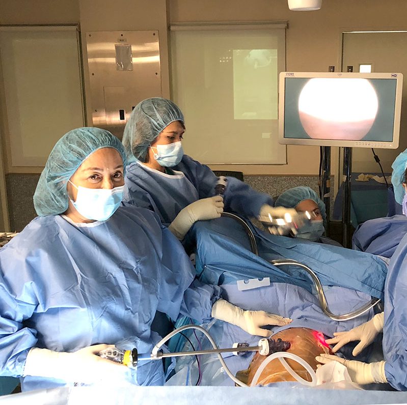
Five years later, Mrs. J. refers an acquaintance, also pregnant for the first time at 16 weeks, with a 7×7 cm. ovarian cyst. By this time, the technology of minimally invasive therapy through laparoscopy had come of age. I convinced the patient that laparoscopic removal was the best to do in this case. Three 1-cm incisions were made on the abdomen above the level of the umbilicus, the mass was visualized, excised and later evacuated by slightly enlarging the right hole where the instruments were being inserted. It was almost like removing a tennis ball through a keyhole.
Intraoperatively and postoperatively, the patient received no tocolytic medications to prevent premature labor. She never manifested with premature contractions post-op. The baby was monitored until the patient was discharged the following day in good condition. The patient and her whole family were extremely amazed and grateful for her small incisions and remarkable post-op recovery.
What is Minimally Invasive Surgery or Laparoscopy?
In non-pregnant women, laparoscopic removal of ovarian cysts involves making a 1-cm incision in the umbilicus, inserting a tube telescopic rod attached to a video camera and a fiberoptic light source. Carbon dioxide gas is then used to blow up the abdomen much llke a balloon to lift the abdominal wall from the intestines and create a working space. This gas is natural to the human body and is later absorbed and eventually removed by the respiratory system. Two other incisions are made on the left and right side of the lower abdomen to insert ports under camera guidance. This is where the instruments like graspers, scissors, etc are inserted into the abdominal cavity. Then, through a tv monitor to which the camera image of the abdominal cavity is seen, the surgery is accomplished with the instruments as a remote extension of the surgeon’s hands.
In pregnancy, the incision is made midway between the umbilicus and the lower tip of the breastbone to provide clearance to the uterus to insure that the pregnancy will not be injured upon entry of the ports and instruments.
WHAT ARE THE ADVANTAGES?
- Dramatically smaller scar, therefore, less post-operative pain and less need for pain medications.1
- Less hemorrhage thus reducing the chances of blood transfusion
- Less chances for wound infection due to reduced exposure of internal organs to possible external contaminants and due to a smaller skin area traumatized and exposed to skin bacteria.
- Less chances of incisional hernias
- Increased chances of early mobilization because of minimal pain. The patient can usually go home the following day or even on the same day.
- Early mobilization also reduces the chances of thromboembolic complications post-operatively
- (formation of a clot (thrombus) in a blood vessel that breaks loose and is carried by the blood stream to plug another vessel either in the lungs (pulmonary embolism), brain (stroke), gastrointestinal tract, kidneys, or leg))
- Conventional surgery for ovarian cysts in pregnancy recommends waiting for the proper window of time to perform an open surgical exploration during the second trimester of pregnancy. This was because there was a reported 12% abortion rate associated with early exploration in the first trimester and up to 40% increased risk for premature labor if surgery is done in the 3rd trimester. In contrast, research has shown that laparoscopic surgery in pregnancy may be done any time in the 1st, 2nd or early 3rd trimester without increased risk from the usual operative risks.3,1,4 Since there is less or no manipulation of the uterus, which contains inside the developing baby. This leads to less uterine irritability which leads to less chances of abortion or premature labor.8
- Due to decreased need for narcotic pain medications post-operatively, there is less chance of depressing the heart rate and placental perfusion and metabolism of the fetus.
- Due to decreased need for narcotic pain medications post-operatively, there is less chance of depressing the heart rate and placental perfusion and metabolism of the fetus
- Unlike conventional surgery where the pregnant patient is placed in a supine position, the gravid uterus places pressure on the inferior vena cava resulting in decreased venous return to the heart. This decrease in venous return results in the drop in blood pressure of the mother, reduced output of the heart by 10% to 30% and decreased flow of blood to the placenta during surgery5,6,7. In performing laparoscopic surgery, the patient may easily be kept in a left lateral recumbent position. This will shift the uterus to the left of the vena cava, thus, improving venous return and cardiac output5.6, 14.
- The patient can go back to work sooner.9,10,11
- No growth or developmental delay was found in eleven children followed up till 8 years after lap surgery on the mother12.
WHAT ARE THE RISKS COMPARED TO OPEN SURGERY?
- All laparoscopic surgeries are done with a double set-up. This means that should there be a complications arising in the course of laparoscopic surgery preventing the surgeon from proceeding, the surgeon may convert to an open surgery.
- The most significant risks are from insertion of the initial trocar since this is usually a blind procedure. There may be blood vessel or bowel injury particularly if the patient has had a previous surgery. Blood vessel injury can result in hemorrhage that may require blood transfusion. Bowel injuries, especially if unrecognized, can lead to delayed peritonitis. 8
- Since carbon dioxide is insufflated into the abdominal cavity to raise the abdominal wall enough to see the abdominal organs. Upward displacement of the diaphragm with air in the abdominal cavity or pneumoperitoneum in a pregnant patient can cause a decreased lung volume and functional capacity, which may possibly impair the delivery of oxygen to the tissues and to the fetus.
- Very rarely, patients have sustained electrical burns unseen by surgeons who are working with electrocautery machines, the electrodes of which leak current into surrounding tissue. The resulting injuries can result in perforated organs and can also lead to peritonitis.
- Patients may experience shoulder pain afterwards which can result from a pocket of CO2 gas rising in the abdomen. This can end up pushing against the diaphragm, putting pressure on the phrenic nerve which can produce a sensation of pain extending to the shoulders and can make breathing very uncomfortable. Luckily, this phenomenon is transient and will disappear once the CO2 is absorbed by the tissues and eliminated through respiration. 13
Advanced technology has made minimally invasive surgery a preferred mode of intervention particularly for removal of benign ovarian cysts. In pregnancy, it becomes even a superior procedure compared to open surgery because of the reduced discomfort for the mother post-operatively the minimal to absence of manipulation of the uterus plus the tinier scars that are left with the patient as a reminder of her surgery during pregnancy.
- Curet, M.J., et al., Laparoscopy during pregnancy. Arch Surg, 1996. 131(5): p. 546-50; discussion 550-1.
- Curet, M.J., Special problems in laparoscopic surgery. Previous abdominal surgery, obesity, and pregnancy. Surg Clin North Am, 2000. 80(4): p. 1093-110.
- Reedy, M.B., B. Kallen, and T.J. Kuehl, Laparoscopy during pregnancy: a study of five fetal outcome parameters with use of the Swedish Health Registry. Am J Obstet Gynecol, 1997. 177(3): p. 673-9.
- Oelsner, G., et al., Pregnancy outcome after laparoscopy or laparotomy in pregnancy. J Am Assoc Gynecol Laparosc, 2003. 10(2): p. 200-4.
- Elkayam U, G.N., Cardiovascular physiology of pregnancy. Cardiac Problems in Pregnancy: Diagnosis and Management of Maternal and Fetal Disease, ed. G.N. U
- Clark, S.L., et al., Position change and central hemodynamic profile during normal third-trimester pregnancy and post partum. Am J Obstet Gynecol, 1991. 164(3): p. 883-7.
- Gordon, M.C., Maternal Physiology in Pregnancy, in Obstetrics: Normal and Problem Pregnancies, S.G. Gabbe, J.R. Niebyl, J.L. Simpson, Editor. 2002, Churchill Livingstone: Philadelphia. p. 63-91
- Janie Fuller, DDS, (CAPT, USPHS), Walter Scott, Ph.D. (CAPT, USPHS), Binita Ashar, M.D., Julia Corrado, M.D. FDA, CDRH, Laparoscopic Trocar Injuries: A report from a U.S. Food and Drug Administration (FDA) Center for Devices and Radiological Health (CDRH) Systematic Technology Assessment of Medical Products (STAMP) Committee, Finalized: November 7, 2003
- Andreoli, M., et al., Laparoscopic surgery during pregnancy. J Am Assoc Gynecol Laparosc, 1999. 6(2): p. 229-33.
- Shay, D.C., K. Bhavani-Shankar, and S. Datta, Laparoscopic surgery during pregnancy. Anesthesiol Clin North America, 2001. 19(1): p. 57-67.
- Oelsner, G., et al., Pregnancy outcome after laparoscopy or laparotomy in pregnancy. J Am Assoc Gynecol Laparosc, 2003. 10(2): p. 200-4.
Elkayam. 1982, New York: Alan R Liss. 5.. - Rizzo, A.G., Laparoscopic surgery in pregnancy: long-term follow-up. J Laparoendosc Adv Surg Tech A, 2003. 13(1): p. 11-5.
- Abdominal pain after laparoscopy: the value of a gas drain. Br J Obstet Gynaecol. 1987 Mar;94(3):267-9
- 65. Clark, S.L., et al., Position change and central hemodynamic profile during normal third-trimester pregnancy and post partum. Am J Obstet Gynecol, 1991. 164(3): p. 883-7.
66. Gordon, M.C., Maternal Physiology in Pregnancy, in Obstetrics: Normal and Problem Pregnancies, S.G. Gabbe, J.R. Niebyl, J.L. Simpson, Editor. 2002, Churchill Livingstone: Philadelphia. p. 63-91. - Reedy, M.B., et al., Laparoscopy during pregnancy. A survey of laparoendoscopic surgeons. J Reprod Med, 1997. 42(1): p. 33-8.
The key element in laparoscopic surgery is the use of a laparoscope. There are two types: 1)a telescopic rod lens system, that is usually connected to a video camera (single chip or three chip) or a digital laparoscope where the charge-coupled device is placed at the end of the laparoscope, eliminating the rod lens system.[1] Also attached is a fiber optic cable system connected to a ‘cold’ light source (halogen or xenon), to illuminate the operative field, inserted through a 5 mm or 10 mm cannula or trocar to view the operative field. The abdomen is usually insufflated with carbon dioxide gas to create a working and viewing space. The abdomen is essentially blown up like a balloon (insufflated), elevating the abdominal wall above the internal organs like a dome. The gas used is CO2, which is common to the human body and can be absorbed by tissue and removed by the respiratory system. It is also non-flammable, which is important because electrosurgical devices are commonly used in laparoscopic procedures.
5-10mm diameter instruments (graspers, scissors, clip applier) can be introduced by the surgeon into the abdomen through trocars (hollow tubes with a seal to keep the CO2 from leaking).